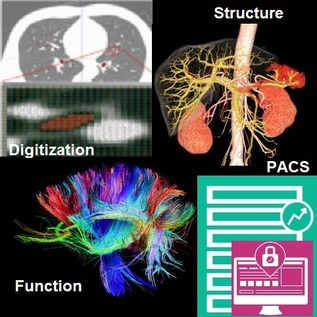
Image digitization has paved the way to effective structural visualization of diseased tissue or organs; today imaging has begun to have implications that transcend merely diagnostic value and is entering the realm of surgical planning minimally- or non-invasive examination and treatment through the realistic depiction of three-dimensional ‘depths’ of medical imaging as it relates to specific anatomical shapes. Image post-processing capabilities embedded in digital image management systems today often amplify the value of visualization by facilitating extraction of two or three dimensional measurements which is useful for purposes of reporting, and may employ cutting-edge digital signal processing technologies that quantify (or semi-quantify) either static or time-series image datasets. This augments the end-user’s cognitive capabilities by serving as a physician’s second-reader to accurately diagnose disease or plan out surgical decisions.
Quantification of images has in-turn led into the concept of computer aided diagnostics (CAD), wherein a physician receives a diagnosis or ‘result’ from a non-human entity. CAD may be dubbed as ‘clinical intelligence’ to support daily radiology tasks and is often based on techniques employing machine-learning and data-based rule-learning technology which actually ‘arrive at’ a clinical decision rather than merely ‘guiding’ a physician towards one. This concept itself germinated in the 1960s but today has matured into a major focus of biomedical and clinical research relating to imaging-based biomarker discovery. Developments in this field of CAD have been incorporated into the routine diagnostic radiology approach to the structural screening of breast cancer on mammograms, early detection of heart disease [3-5] and even estimation of rupture risk of vascular aneurysms, to name just a few applications being investigated in this mushrooming space. But wait… there’s more! We can do even more with imaging today…
One of the research domains receiving the most attention from funding agencies including the National Institutes of Health in the recent past has been the enabling of existing capital equipment with capabilities of imaging neuronal, cardiovascular and cellular ‘function’ as an extension to the convention of visualizing the structure of tissue or an organ. As opposed to structural imaging, functional imaging focuses on revealing physiological activities within a certain tissue or organ by employing medical image modalities that usually reflect a spatial distribution of injected tracers or probes within the body. Functional imaging has probably seen some of its greatest application in cognitive neuroimaging i.e. understanding the link between neuronal activity and functional imaging signals. A few functional imaging modalities which have made an impact in this space, to name a few, include positron emission tomography (PET), infrared imaging, Electroencephalography (EEG), Magnetoencephalography (MEG), functional magnetic resonance imaging (fMRI) [2] to detect blood-oxygen-level-dependent contrast material as an indicator of brain neuronal activity, and diffusion weighted imaging conducted by the Human Connectome Project which aims to understand the details of neural connectivity and build for the first time an integrated roadmap of structural as well as functional neural connections within the brain [3]…
References:
[1] DICOM. http://medical.nema.org/Dicom/
[2] Journal of Neuroscience Methods 74 (1997) 229–243. http://neurosci.info/courses/systems/FMRI/kim_fmri.pdf
[3] http://www.humanconnectomeproject.org/about/


 RSS Feed
