Offered in Fall 2017
Introduction to Biomedical Imaging
BMED 444 / 544: Biomedical Engineering Program, Duquesne University

Instructor: Prahlad G Menon, PhD
Course Description:
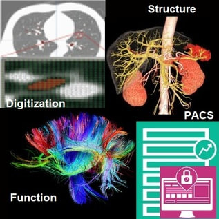
BMED 444 / 544 undergraduate course, open to graduate student enrollment, which introduces the fundamental principles of imaging and image processing from major modalities – X-ray, CT, MRI, Ultrasound and Optical Imaging systems – used in clinical medicine and biomedical research. The course is a combination of lectures as well as demonstrations which introduce the fundamentals of acquiring and processing images from a signals & systems standpoint, grounded on mathematical modeling of imaging systems. Therefore, while the emphasis will be on imparting an understanding of physics behind tomographic imaging devices, the course provides students with some intuition in regard to engineering effective image processing pipelines for visualization or analysis of acquired images.
A strong foundational understanding of imaging techniques will be established through assignments involving simulation of image acquisition processes. To this extent, while the course has no specific pre-requisites, it will involve programming in Matlab (The Mathworks Inc, Natick, MA) for implementation and exploration of the key mathematical concepts.
The course also provides an overview of a wide range of applications of imaging data, including fundamental methods for deriving quantitative biomarkers of disease or disease progression, image rendering / visualization for surgical planning and real-time interventional guidance. Fundamental medical image processing techniques such as image filtering, segmentation & registration are introduced from the standpoint of contextual examples and case studies.
Course Objectives: Upon course completion, students will have a strong fundamental understanding of the physics behind tomographic imaging devices for biological and medical imaging. More specifically, students will be able describe the physics of image formation, build linear systems models and further programmatically simulate image formation of specific imaging modalities relevant to biomedical imaging.
Topics Covered:
- Principles of imaging from major medical modalities
- Image visualization and rendering
- Fundamental image filtering in the time domain and frequency domain
Class Schedule: Class meets twice a week for 75 minutes each day, which will include instructor-guided Matlab programming sessions.
Learning Objectives
Introduction to Biomedical Imaging

his course is designed with the overarching goal of imparting a strong fundamental understanding of the physics behind tomographic imaging devices. While the emphasis is on biomedical imaging, since the course is grounded on mathematical modeling of imaging systems, students will also gain a keen intuition in regard to designing effective image processing pipelines for visualization or analysis of acquired images.
Therefore, the learning objectives of this course are:
• To describe the physics of image formation, build linear systems models of the same and further programmatically simulate image formation of specific imaging modalities relevant to biomedical imaging.
• To build image processing pipelines for image enhancement, feature extraction, image segmentation & registration, and to programmatically implement at least basic image processing filters in both image space as well as in the Fourier domain.
Prerequisites: Basic vector calculus and linear algebra (or permission of the instructor), basic signal processing course (Fourier transforms and 1D signal processing), Programming in Matlab (The Mathworks Inc, Natick, MA) for implementation and exploration of the key mathematical concepts.
Texts & References:
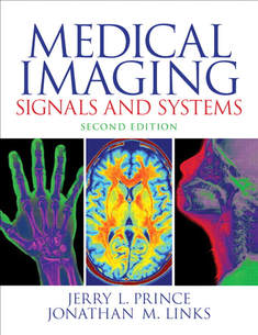
1) Medical Imaging Signals and Systems, by Jerry Prince and Jonathan Links, Prentice Hall, 2nd edition. ISBN-13: 978-0132145183
2) Fundamentals of Medical Imaging, by Paul Suetens, Cambridge Press, Second Edition (2009) ISBN 978-0-521-51915-1.
3) Digital Image Processing (3rd Edition), by Rafael C. Gonzalez & Richard E. Woods. ISBN-13: 978-0131687288.
