Offered in Spring 2015, 2016
Engineering Medical Devices for
Quantitative Image Analysis & Visualization
BIOENG-2385: Dept of Bioengineering, University of Pittsburgh

Instructor: Prahlad G Menon, PhD
Course Description:
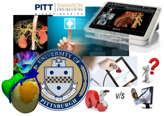
The need for this course stems from an increasing demand for medical device engineers to support a rapidly evolving industry built upon technologies leveraging biological and medical imaging data (including from Optical Imaging, MRI, CT, Ultrasound, OCT and other modalities). This rapidly growing, research-intensive industry supports software based clinical applications which augment diagnostic capabilities of physicians for the early & accurate detection of disease, as well as extend the abilities of surgeons to guiding therapeutic strategy based on image data or computationally modeled pre-surgical plans which forecast patient-specific response under different scenarios.
Students will be familiarized with the image-formation principles of various imaging modalities (eg: Magnetic Resonance Imaging (MRI), X-ray Computed Tomography (CT), Fluoroscopy, Ultrasound and Optical Systems for Microscopy), as well as how the 2D, 3D and 4D (3D + time) data created from these imaging modalities are optimally acquired and processed for quantification and visualization purposes. The course will not only delve into algorithmic implementation details of methods in image processing while providing ample opportunity for students to explore these methods in a variety of feature spaces, but will also train and offer the opportunity for students to piece together complex image processing pipelines based on contemporary software tools and open-source libraries (including, Matlab, OpenCV, SimpleITK, ITK and VTK). Further, emphasis will be laid on the translation of these image-processing concepts and workflows to real-world applications in the clinic, especially in applications relating to improved medical diagnosis, planning of treatment strategy and image-guided surgery. Finally, the relevance of medical device regulation and approval (i.e. FDA, CE-mark clearances etc.) will be highlighted specifically in the context of image post-processing technologies for clinical workflow augmentation.
A special topic which will be covered in the interest of added stimulation for final projects will be “Image Based Computational Biomechanics & Fluid Dynamics” which will include an overview of the methodology to get from medical images to a discrete computational analysis of patient-specific vascular blood flow as well as an interactive lab session on using a commercial numerical solver framework (eg: ANSYS Fluent).
Learning Objectives
Engineering Medical Devices for Image Processing, Biomodeling and Visualization

This course is designed with the overarching goal of training a new breed of biomedical device engineers equipped with a thorough knowledge of both implementation detail of various algorithmic image processing and engineering principles used in the conception, design, development, analysis and operation of innovative biomedical imaging based techniques, and also trained in the translation of such technologies into the clinic for augmenting basic biological science research, image biomarker discovery, medical diagnostics and surgical planning or surgical assist technologies.
The learning objectives of this course are:
• To introduce students the fundamentals of medical image formation, image processing, computational geometry, numerical methods for analysis of medical image data and data visualization and it relates to engineering medical devices ready for clinical translation.
• To shed light on the relevance of medical device regulations on device safety and efficacy specifically in the context of image post-processing technologies for clinical workflow augmentation.
Prerequisites: Basic vector calculus and linear algebra (or permission of the instructor), as well as a working knowledge of Matlab programming.
Text books:
1) Machine Vision, by Wesley E. Snyder & Hairong Qi, © 2004, ISBN 978-0-521-16981-3 (paperback) or 978-0-521-83046-1 (hardback)
2) Insight into Images: Principles and Practice for Segmentation, Registration and Image Analysis, edited by Terry S. Yoo, © 2004, ISBN 1-56881-217-53)
3) Digital Image Processing (3rd Edition), by Rafael C. Gonzalez & Richard E. Woods. ISBN-13: 978-0131687288.
Syllabus & Course Material, Spring 2015
Available upon request via email; write to: prm44@pitt.edu
or via CourseWeb / Blackboard for registered students.
Second opinions and medical technology solutions for the Patient.
We offer timely and accurate image processing of radiology images for clinical care, research, and training.
This is a service brought to you by the MEdical Diagnostics and CArdio-Vascular Engineering Lab.
The MeDCaVE – where QuantMD is engineered.
This is a service brought to you by the MEdical Diagnostics and CArdio-Vascular Engineering Lab.
The MeDCaVE – where QuantMD is engineered.
