• Home
  • The MeDCaVE - Research
  • Teaching
    • Teaching
    • BMED444
    • BMED-310-Bisignals&Systems
    • BMED-110-IntrotoProgramming
    • BIOENG-2385 Spring 2015
    • BIOENG-1330-2330
    • BIA Fall 2014
    • Engineering Pittsburgh's High School Curriculum
    • Communicative English
    • Prospective Student?
    • Passing Thoughts
  • Students
    • Students-NoLinks
    • Graduated Degree Students
  • Press
    • Community outreach
    • The MeDCaVE / QuantMD Press
    • FF2Israel2014
    • CIRC Review May 2014
  • Personal
    • The Menon Blog
    • Fiverr.com Gigs
    • Need a 2nd Opinion..?
  • Contact me!
    • Contact me!
    • Jobs
Prahlad G Menon, Ph.D Associate Professor -The MeDCaVE Lab

The Cornerstones of a Radiology Practice in the Digital Era

5/23/2014

33 Comments

 
Picture
Print radiology systems are inherently inefficient and often of poor quality, therefore making them unsustainable. Owing to both the slowness of image preparation and diminished quality of images, a print-based radiology practice invites a higher rate of professional second opinions and unnecessary re-scanning. This is not only a waste of time that could be spent on managing more patients but such inefficiencies also present a financial drain on the healthcare system. The solution to these shortcomings is the use of digital radiology systems.  However, in order to truly appreciate the impact of digital systems in the radiology practice, one must first understand how the individual component systems of a digital radiology practice operate. A digital practice primarily involves two key components - radiology information systems (RIS) and picture arhiving and communication systems (PACS). This article provides a high-level overview of these component systems as well as how they have together revolutionized the radiology practice.

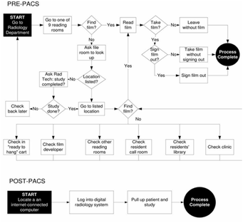
Prior to the implementation of the digital systems in radiology practices, studies have indicated that physicians spent an average of one to three hours ‘searching’ for hard-copy films during the day [1].  Shown below is a flow-diagram illustrating a typical hospital workflow for reviewing radiology images (adapted from [1]) in the pre-digital (or pre-PACS) era as opposed to reviewing images on a digital system (namely, PACS).  This illustration helps one truly appreciate the inefficiencies of a pre-digital radiology practice, specifically in terms of time spent developing, retrieving and interpreting radiology images. Digital data storage and retrieval eliminates several steps in an otherwise convoluted print pipeline, paving the way for real-time reporting, on-the-fly quantitative analyses and minimal paper-pushing.  The digital age and its manifestation in radiology practice has helped lower costs of radiology practices the world over, while increasing efficiency and quality of reporting.

The RIS is a computer system designed to support operational and business workflows within a radiology department. It is a repository of patient data and report which is often populated into the electronic patient record. However, an RIS by itself is debilitated in the capability to store and access the radiology images themselves!  This is the role that PACS fills in; while the RIS manages patient data and department scheduling task, PACS specifically focuses on images. PACS is in principle a three-component assembly integrated together by digital networks which is constituted of an image data acquisition gateway (i.e. connections to the imaging systems themselves!), a server with substantial data-archival facilities, and of-course several display workstations for retrieving, reviewing and reporting on the acquired and stored images. An RIS integrated with a PACS makes patient data and image access lightning-fast for busy physicians whom shouldn’t be spending inordinate amounts of time on data or image access.

While an RIS integrated with a PACS, like any digital system, could certainly fall prey to the known risks for technological disaster such as unforeseen power-failure or data-corruptions in storage media, their advantages greatly outweigh such low-likelihood adverse events. In any event, modern PACS are equipped with the ability to recover from such disasters through programs that regulate routine data-backups and disaster recovery, reasserting the fact that digital data storage and retrieval is quite a reliable system.  Further, digital image storage through direct connectivity with source imaging acquisition systems (eg: X-Ray, MRI, CT etc.) drastically diminishes the chance that images are “lost in transit”, presenting another major advantage over print-based radiology.

To conclude, RIS and PACS – the cornerstones of today’s digital radiology era – have together made a tremendous impact on the overall sustainability and quality-of-care offered by modern radiology practices.  PACS-empowered digital radiology practices have great potential to further augment quality of care through integration with quantitative image post-processing software packages, therefore making them an infinitely extendable platform technology equipped to catapult your radiology practice into the future of quantitative imaging based healthcare.

References:

[1] Srinivasan, M. (2012). Saving time, improving satisfaction: the impact of a digital radiology system on physician workflow and system efficiency. Digital _Medicine, 1(1).


33 Comments
Forward>>

    Personal thoughts on Imaging, IoT, Megatrends, Technology & Travel - 
    The "New" Menon Blog

    Picture

    Prof. Prahlad G Menon, PhD

    Dr. Menon is an Associate Professor of Mathematics with appointments in Bioengineering at University of Pittsburgh and Biomedical Engineering at University of Texas at San Antonio.  He was previously a tenure-track, early-career assistant professor with the department of biomedical engineering at Duquesne University (Pittsburgh, PA) and until May 2015 on the faculty of the electrical and computer engineering (ECE) department in Carnegie Mellon University joint institute of engineering with Sun Yat-sen University (Pittsburgh, PA, USA and Guangzhou, China), where he currently maintains an adjunct professor appointment. He has served as adjunct faculty with the Dept of Biomedical Engineering at Carnegie Mellon University as well as the Heinz College of Information Science at Carnegie Mellon University. Dr. Menon's research group, The MeDCaVE, has its interests in the broad area of AI / data science applied to medical imaging analysis for biomarker discovery and more specifically in computational simulation of vascular flows and cardiovascular biomechanics, with application to diagnostics, surgical planning and interventional guidance.

    Also see the Prospective Student blog if you are a prospective student wishing to become affiliated with The MeDCaVE research group.


    Archives

    March 2023
    November 2020
    September 2017
    August 2017
    May 2016
    February 2016
    December 2015
    November 2015
    February 2015
    October 2014
    August 2014
    July 2014
    June 2014
    May 2014

    Categories

    All
    Chess
    Cloud Computing
    Condition Based Monitoring
    Daily Price Predictions
    Daily Price Predictions
    Data Science
    Education
    Healthcare
    IBM Bluemix
    IoT
    Market Sentiment Analysis
    Market Sentiment Analysis
    Medical Imaging
    Megatrends
    Nature
    Outreach
    Pacs
    Poetry
    Predictive Maintenance
    Radiology
    Real Estate
    Social Work
    Stock Market
    Stock Market
    Supercomputing
    Travel

    RSS Feed

Copyright 2012-14, Prahlad G. Menon
Free Domains